Gene therapy vectors: Preclinical testing with retinal organoids
A recent collaborative study with Professor Robert MacLaren from the University of Oxford, validates the use of retinal organoids as an in vitro model for the evaluation of new gene therapy vectors for retinal diseases.

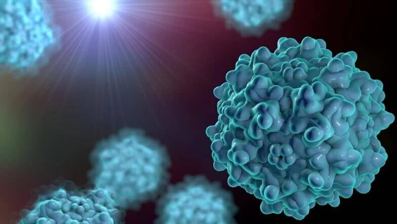
Adeno Associated Virus (AAV) vectors have emerged as the most successful retinal gene therapy vectors following the FDA approval of Luxturna in 2017. This new gene therapy product is now used to treat blindness caused by mutations in the RPE65 gene in the retinal pigment epithelium (RPE), a thin cell layer at the back of the eye that support and nourishes the light-sensing cells or photoreceptors. As most common retinopathies cause blindness due to the deterioration of photoreceptors or retinal pigment epithelial cells (RPEC), gene therapy vectors need to target these cells to deliver the therapeutic gene and correct the defect. Further improvements are required to improve the transduction efficiency of photoreceptors by viral vectors. Carefully engineered modifications of the viral capsid of AAV2 enhance transduction of photoreceptors and show significant improvement of gene delivery. Examples such as AAV serotypes 2 and 8 with a small peptide insertion (AAV2 7m8) or with single amino acid substitutions of exposed tyrosine residues of the capsid (AAV8 Y733F, AAV2 Y444F and AAV2 quad Y-F) are likely to improve gene delivery into photoreceptors1 in patients. Although some of these AAV serotype variants have been tested in rodents and dogs, there is insufficient evidence relating to their transduction efficiency in the human retina highlighting the need to develop in vitro models that recapitulate the human eye.
Newcells’ has developed a retinal organoid model based on human biology. The organoids are iPSC-derived, recapitulating retinal development and thus mimicking the structure and function of the human tissue. Our recently published work, validates their use for screening and selection of optimal AAV vectors for gene therapy. By testing a range of AAV vectors composed of different capsid variants, transgenes and promoters using retinal organoids, we were able to select the most optimal to transduce the desired human retinal cells. Given the limited availability of human in vitro and ex-vivo retinal models, human retinal organoids represent a uniquely valuable in vitro model for screening. The results reveal robust transduction of photoreceptor-like cells and demonstrates the safety of the AAV vectors used. These results pave the way to accelerated development of novel gene therapies for retinal diseases.
The results reveal robust transduction of photoreceptor-like cells and demonstrates the safety of the AAV vectors used. These results pave the way to accelerated development of novel gene therapies for retinal diseases.
Newcells’ novel iPSC-derived human retinal organoids are light-responsive and validated. It takes 150 days in culture and expert knowhow to generate and characterise these organoids. The retinal organoids are complex but with the help of Newcells’ experts, they become a robust in vitro model for the evaluation of new gene therapy vectors for retinal diseases. They can be delivered to your laboratories for in-house studies or alternatively, the work can be outsourced to Newcells. Our service includes the design, development, and completion of study protocols in our UK laboratories, working closely with our customers to define a tailored study design to answer specific scientific queries.
Why not read the paper recently published in collaboration with Professor Robert MacLaren at the University of Oxford.
Interested to know more about the use of retinal organoids in preclinical testing of AAV vectors?
Contact usShare on social media:
Don't miss out on our latest innovations: follow us on Linkedin
Newcells Biotech
15th June, 2022
Retina
Update


